June 10, 2017

Get to know Phycomyces blakesleeanus

Get to know Phycomyces blakesleeanus


Phycomyces blakesleeanus are fungi that filamentous and known to be very sensitive to their environment including light, wind, gravity and objects around them. 

They are known to have phototrophic growth and they mate with their opposite "sex". They exist in two different forms the (+) and (-).

Their optimal growth takes place in potato dextrose medium between 25 and 30°C. 


Under light microscope
On petri dish culture


For information check out:

https://www.wikigenes.org/e/mesh/e/9666.html

Get to know Sordaria fimicola

Get to know Sordaria fimicola

Sordaria fimicola is a fungus very well known for its usage in studying genetics thanks to its life cycle that respects the Mendelian laws of segregation. It is known to be a haploid organism. For most of its life cycle it is a non-motile organism.

First of all, this fungus grows at 25°C on nutrient agar. In nature these microbes can be found in the feces of organisms that eat plants. 

https://upload.wikimedia.org/wikipedia/commons/6/61/Morelasci.jpg

Why are they used for genetic studies ?

The fungus does sexual reproduction which undergoes mitosis and meiosis. This organism, even though, begins life as a single celled organism it starts its mitotic cycle very early. 

As we know this mitotic division gives birth two identical daughter cells which are haploid. During this process of cell duplication a new "organ" starts growing: mycelium, which is known to be the sexual "organ" of the fungus. When the mycelium of two organisms meet, there is birth of diploid zygote. This zygote enters meiosis which ends up with the birth of 4 daughter cells also known as spores. The zygote does meiosis twice which allows the formation of 8 ascospores. This a linear organisation of the eight spores which are contained in a bag called asci. 

There are two alleles which determine whether as ascospores are white or black. This allows us to study the different genetic process taking place in simple organisms.

For more information check out:

http://botit.botany.wisc.edu/toms_fungi/mar2007.html

Get to know Micrococcus luteus

Get to know Micrococcus luteus

The Micrococcus luteus  is known as an actinobacterium that has one of the smallest genomes in bacteria. The bacterium is between 0.5 and 3.5 micrometers in diameter. Its genome size has been identified to be around 2,501,097 bp in a single circular chromosome. It can be found in various environments such as water, soil, dust and even human skin. 

This bacterium is also used in quality control for sterility testing and asseying antibiotics and fungicides. 


Figure 1: M.luteus in nutrient agar medium as received from the supplier and conserved in the fridge
This bacterium can be grown on nutrient agar or broth medium. For the recipe for this medium look at the previous post "Get to know Pseudomonas fluorescens". 

However we wanted to know, if this microbe could grow on a medium that is very close to the nutirient medium in composition, the LBA medium. In order to test the growth of the M.luteus in LBA medium, we did a overnight culture of the latter. As expected it did grow in the LBA medium.


The optimal growth temperature for this bacterium is between 25°C and 30°C.

This cocci is also known as nonmotile and clustered. Furthermore these bacteria produce a yellowish water insoluble pigment which has been identified as a carotenoid. This pigment is said to absorb wave-lenghts from 350 to 475 nanometers.

For more information check-out:

https://microbewiki.kenyon.edu/index.php/Micrococcus


Quorum sensing: Bacterial communication


What is quorum sensing ?

Quorum sensing (QS) is a communication system used by bacteria to communicate between each other in an environment. Bacteria produce specific molecules called autoinducers and release them in the environment to communicate with the other cells in the same environment.

This communication happens when the cell density high enough. In this case the autoinducers bind to specific receptors of the bacterial cells and induce the transcription of genes involved in pathogenicity, bioluminescence, biofilm formation and antibiotic production for example.

Scientist are working on quorum sensing to replace the traditional antibiotic treatment of bacterial infection. By targeting the communication between bacteria instead of killing them,we might be able to overcome bacterial resistence.

What did we do ?

We learned that some spices we use in our day to day life are effective in ihibiting quorum sensing in some bacteria. Following this, we wanted to know if the these spices had any effect on the bacteria we had in the lab. Basically we wanted to study the effect of basil, cinnamon and ginger on the quorum sensing activity of 
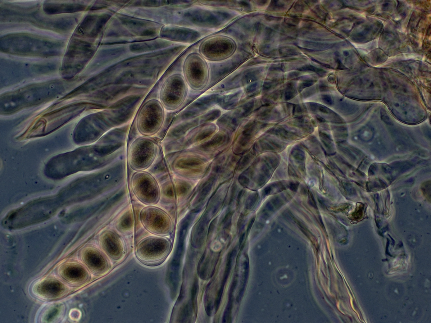
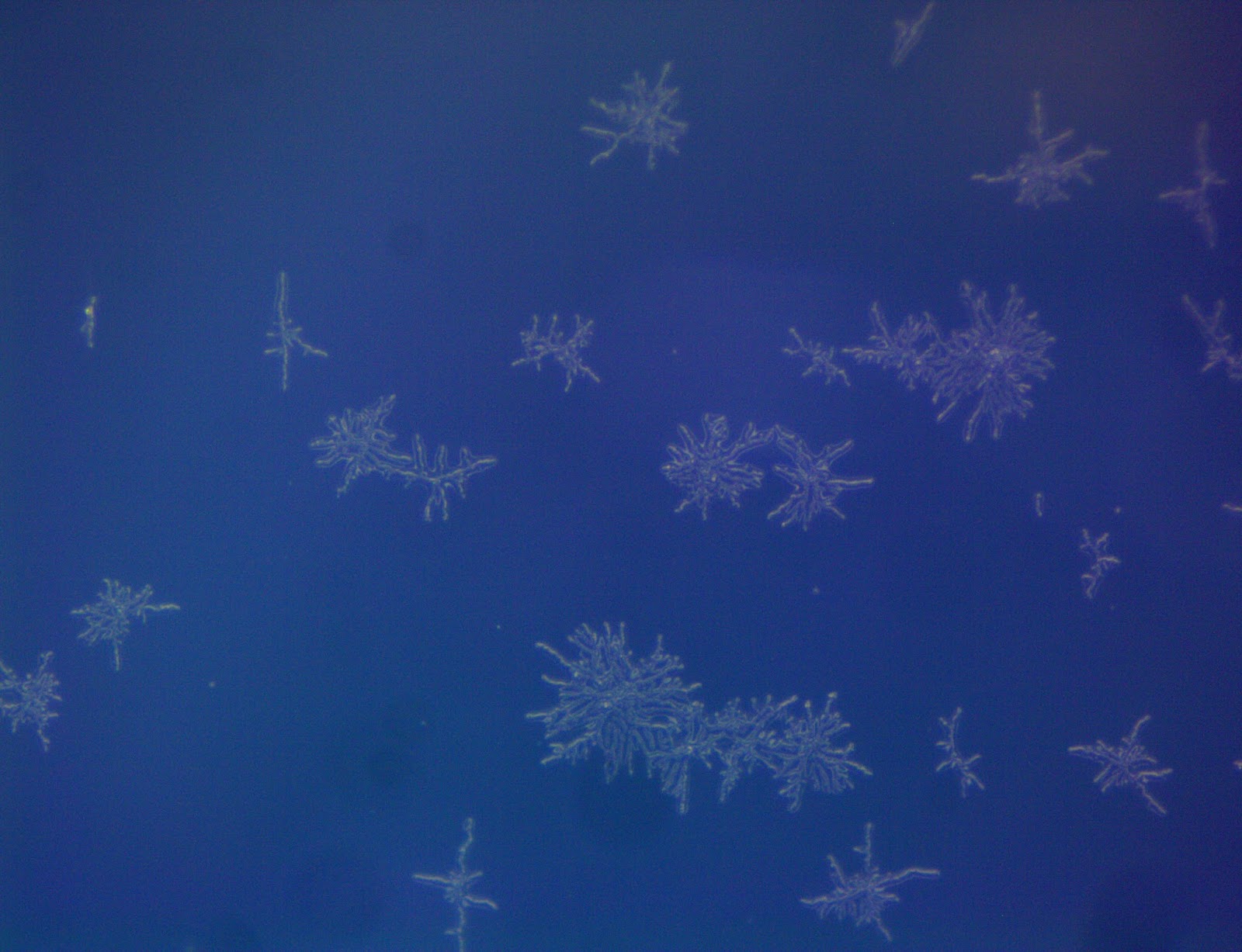
Bacillus megaterium, Escherichia coli, Pseudomonas fluorescens,Vibrio fischeri and Bacillus subtilis. Each bacteria has its own way of quorum sensing. For example, in E.coli  the QS is detected by bioluminescence, biofilm formation and swarming. In bacteria like the V.fischeri  the QS activity is bioluminescence.

Our project was to study the effect the different essential oils had on the QS activity.

In this post I'm going to focus on how the QS activity of V.fischeri was affected and how we studied and analyzed the fluorescence of these cells.

How did we grow the bacteria ?

The experimental setup was the following: the V.fischeri was put in 96-well plate along the other bacteria that we studied. The concentration of essential ils varied from 1% to 0.1% mixed in the media. The samples from the each well was observed on fluorescence microscope using different filters (GFP and DAPI).

Vibrio-trans-GFP-DAPI-63x.tif
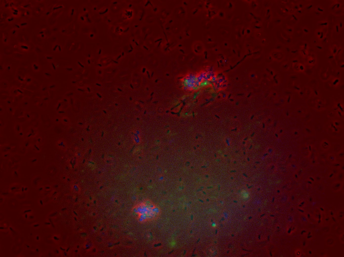
Figure 1: V.fischeri seen under fluorescent microscope with the combination of DAPI (blue) and GFP (green) filters


How was the fluorescence studied ?

We calculated the Corrected Total Cell Fluorescence (CTCF) using Image J for each sample: Photobacterium medium and ginger essential oil at 1%, photobacterium medium and cinnamon essential oil at 1% and finally the control which was only the photobacterium medium.

In order to calculate the CTCF using Image J, we followed the following steps:
- We selected the first cell we wanted the CTFC from. The using any drawing tool from Image J.
- Then in the Analyze menu, we clicked on "set measurements" where we selected AREA, INTEGRATED DENSITY and MEAN GRAY VALUE.
- We clicked on "Measure" from the analyze menu and a window will show up with the different values we asked for the first cell selected using the drawing tool.
- Then we repeated this step for ten cells selected "randomly" (the most random possible)
- In order to take out any background noise, we selected a region from the background without any cells and obtained the same values. This was done 5 times.
- Then we selected all the data and pasted it on an excel sheet.
- The CTCF was calculated using this formula:

CTCF= Integrated density-(Area ×Mean grey value)

The mean CTCF was used for the graph which follows.


The results suggest that when the bacteria grew in a medium with ginger or cinnamon essential the GFP fluorescence (green) decreased drastically compared to the control. The DAPI is also quiet low but doesn't show a high decrease as GFP.

We can conclude little, as no statistical tests were done. However we can see a important impact on these cells when they grew on media with essential oils. Further tests need to be done in order to conclude. But this project can be used as a starting point for further research on this.

For information, check out:

Our google drive:  https://drive.google.com/open?id=0B8RC6xPQ6yh_dkFpS0lJMllrV2s


Phlyctochytrium acuminatum


Latin Name: Phlyctochytrium acuminatum


Common Name: doesn't have a very specific common name

Optimum Temperature: 25 °C

Medium: Emerson YpS (agar surface must be wet!!)
What is it?? FUNGUS

Source: Naiane Rios (click on the picture and zoom in!)

Descriptions:
Posterior flagellated zoospores.
Produces apophysis and rhizoids within host wall


How to prepare the medium Emerson YpS?


Preparation

1) Suspend 40.5 g in 1L of distilled water. If desired, half strength medium can be prepared using 20.25 gr in 1L of distilled water.
2) Heat to boiling to dissolve the medium completely.
3) Sterilize by autoclaving at 15 lbs pressure (121°C) for 15 minutes. 
*Final pH at 7

Posteriorly flagellated zoospores; produces apophysis and rhizoids within host wall

More technical details about the purchase: http://www.carolina.com/fungi/phlyctochytrium-spizellomyces-dolichospermus-living-tube/156170.pr?question=Phlyctochytrium+ 

June 7, 2017

Penicillium notatum


Latin Name: Penicillium notatum

Common name: Penicillium (doesn't have a very specific common name)
Optimum Temperature: 25 °C
Medium: PDA 
What is it?? FUNGUS

Appearance under the loupe: snow flakes!
Source: Naiane Rios (click on the picture and zoom in!)


It goes without saying that Penicillium is mostly known as the source for penicillin and the great medical revolution it provoked.
But the great thing about Penicillium notatum is that it's one of the most easy identifiable fungus there is.

If you see something that has a velvet texture and has a gray-green kind of color, it's very likely you're looking to a pretty Penicillium. You almost wish you could saw from it a long fancy Grammy-Awards-kinda coat. 
Source: http://www.polyvore.com

 How to prepare the medium PDA?
Home Made PDA
 
Preparation
1) To prepare potato infusion, boil 200 g sliced, unpeeled potatoes in 1 liter distilled water for 30 min.  
2) Filter through cheesecloth, saving effluent, which is potato infusion (or use commercial dehydrated form).  
3) Mix with Dextrose, Agar and Water and boil to dissolve
4) Autoclave 15 min at 121°C. 5) Final pH, 5.6 ± 0.2.
Commercial PDA
 
Preparation
1) Add 39 g of the powder to 1 Liter of distilled water. 
2) Boil while mixing to dissolve. 
3) Autoclave 15 min at 121°C
 Macroscopy
This is how Penicillium notatum looks likes under the loupe.
How awesome is that? <3

 Source: Naiane Rios
This is the loupe used, but to have such a cool picture the trick is to have a microscope digital camera attached. The one there is a AmScope MU1000.
 Source: Naiane Rios
Microscopy 
Under the microscope things a bit different.
Expectations: 
Source: Guclu et al., 2010 

Reality: 
Source: http://oldblockwriter.blogspot.fr/


More technical details about the purchase: http://m.carolina.com/fungi/penicillium-notatum-living-plate/156157.pr?question=Penicillium+notatum http://himedialabs.com/TD/M773.pdf

Blog disclaimer

The content created by the Learning thru research Student Bloggers and those providing comments are theirs alone, and do not reflect the opinions of Centre de recherche interdisciplinaire, University Paris Descartes or any employee thereof. The authors of posts and comments are responsible for the accuracy of any of the information supplied on this blog, as well as for any content copyright issues.