
DARK SLIME MOLD
The slime mold Physarum polycephalum(literally meaning: the many-headed slime) is a lover of darkness. It tends to avoid light and to grow in moisture and humidity. It’s bright yellow color makes it particularly interesting to look at, and it’s ability to grow in short time periods is convenient in labs for studying protists and other slime molds. After learning a bit about the organism and it’s photophobia, we wondered how photophobic the slime mould would be if we lit it up several times with different light intensities; so we designed an experiment to do just that!
 The experiment
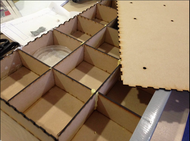
To begin, we built a large box that had 16 compartments in it, each large enough to hold a Petri dish. It was made using a laser cutter, glue, patafix, and silver duct tape.
After this, we prepared our Petri dishes with Agar and water, as well oats at the center. We then placed one oat covered with slime mould in the center of each dish, then used the duct tape to cover half of each plate before placing them in the box. 
We then passed an LED through the top of each compartment to light the plates.
Using an arduino, we set the brightness at which each LED would be powered and also built temperature and light sensors to make sure the conditions didn’t change to much inside the box.
We had 9 plates lit with 3 different intensities, as well as six plates without tape that were either kept in complete darkness or lit at maximum intensity. A final plate was kept just to cultivate the mold.
We expected to see that P. polycephalum would grow and cover a larger portion of the dark side of each plate(under the tape), rather than the portions that were lit by a LED.
Data and data … We analysed them in many way, look!
After one day of growth, we opened the box to make sure that growth had truly taken place and our observations were encouraging, the organism had moderately grown, so we decided to leave our plates another 24 hours of growth. So that’s after more 44 hours of growth that we opened the whole box and took pictures of the plates. Then we measured the area occupied by the organism on each side of the plat (the side that was in the dark and the one that was illuminated).
We compared surface
Once we finally uncovered our boxes, untaped them and took pictures of them, we used the software Image J to see the surface that our slime had covered. Our data didn’t show any clear trend, perhaps we should have the experiment more times. However, at our maximum intensity, the slime mold did seem to flee from the light more than previously, except in one case.
Then we calculated branches
When we actually looked at the plates, we thought that the mold that had grown in the light seemed to be less dense and have less branches, which led us to count the branches on each side of every plate. This turned out to be difficult and unreliable because the quality of our photos was limited.
Finally we calculated fractal dimension

To avoid this difficulty, we decided to calculate the fractal dimension of the P. polycephalum that had grown in the light and under the tape with the same software imagej. To do this, we had to convert the images we had taken to an 8-bit image(in black and white), and then to a binary image, where everything is either entirely black or entirely white.
With the results we built those graphs that show the fractal dimension for the dark place and the the place lit up..

We found a fractal dimension for each value that hardly changed, and as you can see, there are no clear difference between the mold under the tape (in green on the graphs) and under the light (in yellow on the graphs). This meant we could not discover anything about the shape and growth of the mold. They seem to be the same under the light and under the tape.
In contrary that we found in the literature we did not see a difference of growth of the P. polycephalum in the dark or in the light. One of the reason we did not find like the scientific paper can be some problem of our experiment. The quantity of organism we put on the plates was not the same in each plate. Also the limit of the experiment was the lack. But thanks to the measures of the arduino sensors (Temperature and luminosity) we can say that this two parameters did not change and did not disturb the P. polycephalum growth.
If you want to know more check this out :
OUr gitHub: https://github.com/learningthruresearch/Biosensors2017/tree/master/fisarumo
Our Storify: https://storify.com/azpiration/biosensors-final-project
OUr gitHub: https://github.com/learningthruresearch/Biosensors2017/tree/master/fisarumo
Our Storify: https://storify.com/azpiration/biosensors-final-project
- A video : Deep Look. This Pulsating Slime Mold Comes in Peace (Ft. It’s Okay to Be Smart) | Deep Look, 2016. https://www.youtube.com/watch?v=Nx3Uu1hfl6Q.
- Starostzik et al. “A Photoreceptor with Characteristics of Phytochrome Triggers Sporulation in the True Slime Mould Physarum Polycephalum.” FEBS Letters 370, no. 1–2 (1995): 146–48.
- Ueda et al. “ACTION SPECTRA FOR SUPEROXIDE GENERATION AND UV AND VISIBLE LIGHT PHOTOAVOIDANCE IN PLASMODIA OF Physarum Polycephalum.” Photochemistry and Photobiology 48, no. 5 (1988): 705–9.
- Hato et al. “Phototaxis in True Slime Mold Physarum Polycephalum.” Cell Structure and Function 1, no. 3 (1976): 269–78.
- Purschwitz et al. “Seeing the Rainbow: Light Sensing in Fungi.” Current Opinion in Microbiology, Growth and devlopment, 9, no. 6 (2006): 566–71.
- Kakiuchi et al. “Light Irradiation Induces Fragmentation of the Plasmodium, a Novel Photomorphogenesis in the True Slime Mold Physarum Polycephalum: Action Spectra and Evidence for Involvement of the Phytochrome¶.” Photochemistry and Photobiology 73, no. 3 (2001): 324.
- Starostzik. “A Photoreceptor with Characteristics of Phytochrome Triggers Sporulation in the True Slime Mould Physarum Polycephalum.” FEBS Letters 370, no. 1–2 (August 14, 1995): 146–48.
- Deep Look. This Pulsating Slime Mold Comes in Peace (Ft. It’s Okay to Be Smart) | Deep Look, 2016. https://www.youtube.com/watch?v=Nx3Uu1hfl6Q.
- BioFilmer86. Physarum Polycephalum Eating Wild Fungus, 2012. https://www.youtube.com/watch?v=CqkISTDtDJI.
- “Light Sensor (TSL2561).” Accessed February 6, 2017. http://fritzing.org/projects/arduino-light-sensor-tsl2561
No comments:
Post a Comment